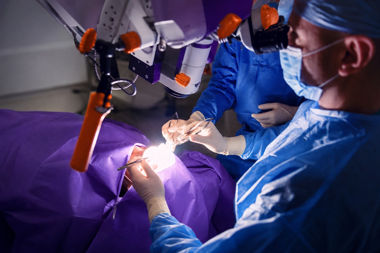
Surgeons in London removed a pituitary tumor while an AI system watched the operation live and marked where hidden blood vessels and nerves were most likely to lie. A camera passed through the patient's nose to the base of the skull fed video to the tool, which tracked the surgical instruments in real time and shaded the areas where the tumor could be cut away more safely. The surgeons kept full control of the procedure throughout. The patient, Hibbert, said afterwards that his sight had come back far enough for him to see the whole room clearly.
The pituitary sits at the base of the brain, beside the arteries that supply it with blood and beside the optic nerves. By The Guardian's account, a one-millimeter error in that space can cause blindness, a stroke or death. This tumor added to the difficulty: it lay deep and out of direct view, so the surgeons had to plot an exact route to it without damaging brain tissue on the way in. That approach phase is where the tool was useful.
It was trained on hundreds of videos of pituitary tumor removals. In preparation, researchers outlined the vessels and nerves by hand on every one of those videos so the system could learn to place them more precisely. Sophia Bano, associate professor of robotics and AI at University College London and the project's technical lead, said the set of examples let the system see far more variations of the operation than a surgeon typically encounters across many years of practice.
The loop inside the operating room is narrow: video in, instruments tracked, probable positions of concealed vessels and nerves marked, safer working zones highlighted. The BBC compared the approach to facial recognition turned on anatomy nobody can see. Hibbert made the case for it in terms of patient safety, noting that there are no road signs inside a head to show surgeons the right way.
What is described here is a capability, not a result. The system reports where vessels and nerves are most likely to be, and "most likely" carries a lot of weight in a space where a millimeter decides between sight and blindness. Nothing in the account states how often the highlighted safe zones were correct, how often they were not, or whether the overlay changed a single decision the surgeons would otherwise have made. For Hibbert the outcome was concrete: he said the operation gave him his life back. That is a fact about a patient, not a fact about a tool.
The quieter part is the annotation. Hundreds of recorded operations were traced by hand, vessel by vessel and nerve by nerve, by people who already knew what they were looking at. That labor is the real asset in this project, and it is the part that does not transfer. The pituitary is one procedure. Every other operation worth this treatment starts from an empty dataset and the same years of manual tracing, which is a far less exciting sentence than the one about hundreds of videos.
Physicians already use AI for clinical notes and symptom lookup, and those uses are forgiving of error in a way this one is not. Pituitary surgery turns on deep knowledge of one patient's particular anatomy, the kind a surgeon builds by studying that person's brain scans in detail beforehand. Experts cited in the reporting worry that doctors will come to depend on AI tools too heavily, and they worry most about the incoming generation of medical students, who will be working alongside such systems from the start of their training.
That is the tension this case leaves behind rather than resolves. The system's competence comes entirely from hundreds of operations performed by surgeons who learned the anatomy without it, by reading scans and building a mental map of one skull at a time. The trainees who inherit the overlay will be learning that anatomy with the answer already drawn on the screen, and the supply of surgeons capable of correcting the tool comes from exactly that group.