
A decade-long collaboration led by the HHMI Janelia research campus, with Google Research and academic partners, has produced the complete wiring diagram of a male fruit fly's central nervous system: 166,000 neurons and 125 million synaptic connections. The work, published as "Sexual dimorphism in the complete connectome of the male Drosophila central nervous system", covers the central brain, the optic lobes and the ventral nerve cord. Because a female map already exists, neuroscience now has two complete nervous systems of the same species that can be laid side by side.
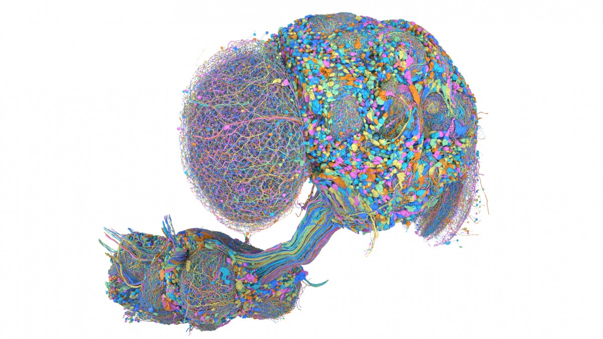
A colorful, highly detailed three-dimensional visualization showing the neurons of a fruit fly's brain and nerve cord
Source: research.google
The ventral nerve cord is what separates this reconstruction from what came before. That structure is the fly's counterpart to a spinal cord, so the map no longer stops at the brain — it describes how the brain drives the body. The central brain, shown in green, the optic lobes in purple and the nerve cord in blue are joined in a single volume, which is what allows a researcher to follow an auditory, visual or olfactory signal through to the motor response it produces. Janelia specialists checked and annotated the result by hand, and the map can be browsed, explored and downloaded in Neuroglancer, Google's open tool for large multidimensional datasets.
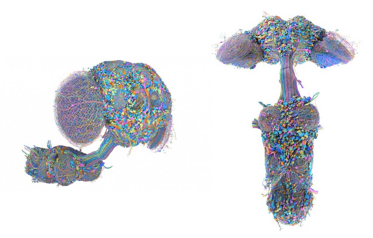
Two detailed color three-dimensional visualizations of the nerve pathways and nerve cord of a fruit fly
Source: research.google
Drosophila melanogaster is an old workhorse of biology, behind discoveries marked by several Nobel Prizes, and its predictable behavior and short life cycle made it a genetics model long before it became a neuroscience one. The reason it is being mapped now is arithmetic: the human brain holds 86 billion neurons, mapping it whole is out of reach, and so the field works on organisms where the full graph is finite.
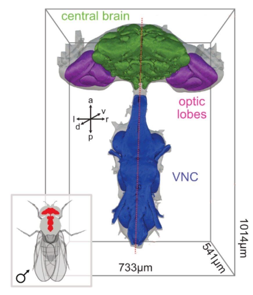
Diagram of the male fruit fly's nervous system showing the central brain, the optic lobes and the VNC
Source: research.google
The male map joins the earlier female brain map and the complete female brain-and-nerve-cord map that followed it. Having both sexes is the point: researchers can compare the regions where neurons differ and study the biological machinery of courtship and aggression, and in the regions built alike in both sexes, two complete maps become a way to study how one individual differs from another. One example already flagged is a neuron present in both the male and the previously mapped female with a different structure in each — the male version carries two extra processes. AI is what recovers the three-dimensional shape of such cells precisely enough for a difference like that to count as a finding rather than an artifact.
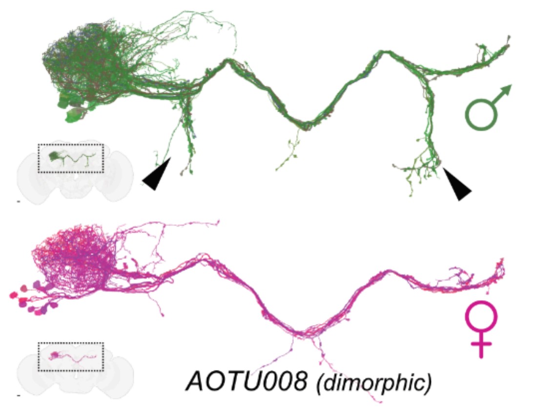
Comparison of the dimorphic AOTU008 pathway showing the differences between male and female fruit flies
Source: research.google
The pipeline behind all of this is unglamorous. A brain is cut into millions of thin slices, every slice is photographed, and computers reassemble the flat electron-microscope images into a three-dimensional reconstruction. Google's tools use flood-filling networks, which start from a single pixel and work out every other pixel belonging to the same object. An earlier, fully automatic reconstruction of the female fly brain came first; then Google and partners released a human-verified map of half the female brain, with 25,000 neurons and 21 million connections, a record at the time. The male map is roughly six and a half times that many neurons and six times that many connections, and it adds the wiring to the body.
The number worth watching, though, is not 166,000. It is how much of this still needs a person. Verifying and describing neuron shapes in a fly brain currently takes years of manual work, and the stated payoff of better automation is that groups could attempt larger mapping projects within the budgets and schedules they actually have. That is an unusually direct admission that the binding constraint is labor, not imaging and not compute. The most recent improvement — adding synthetic neurons to the training data, which made PATHFINDER, the team's self-described most advanced reconstruction system, faster and more accurate — arrives with no number attached to either word. The size of that gain is the figure that decides what is reachable next, and it is the one the announcement does not give.
The 86 billion figure does similar work. It appears as the horizon, and the distance to it is a factor of about 500,000. Read plainly, invoking it alongside a fly is scale-setting rather than a roadmap, and the authors concede as much: a full human reconstruction remains unattainable. What the fly is actually good for is narrower and more defensible — principles of circuit operation, and the processes implicated in Alzheimer's, depression and schizophrenia.
The move that matters more is toward vertebrates, animals with a spinal cord, whose anatomy, evolution and function sit closer to ours. In a Columbia-led study published in Nature, the Google team helped map part of the hindbrain of the elephantnose fish, a region involved in signal processing; that paper, "Connectome analysis of a cerebellum-like circuit for sensory prediction", was the first to show how a static connection map can be combined with other data to study neuroplasticity and learning, producing the most complete mechanistic account of learning in a vertebrate brain to that point. Zebrafish larvae are among the few vertebrates whose brains can be mapped in full today, because at the larval stage they are transparent and neural activity can be measured during the experiment itself — the basis of the ZAPBench dataset. A forthcoming Google and Harvard paper, "A connectome resource for cataloging neurons and analyzing circuits in the larval zebrafish brain", presents the first whole-brain vertebrate dataset that includes both neuron structure and molecular cell types, along with a preliminary release pairing activity and structure in the same larva, open to the research community.
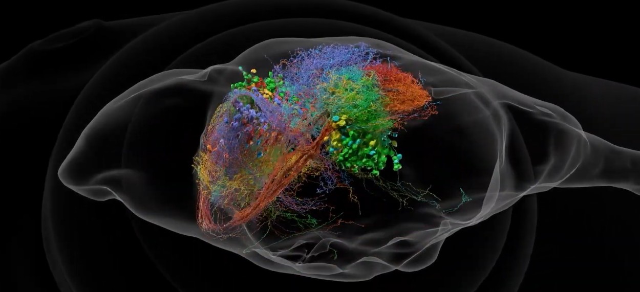
A multicolor three-dimensional map of interconnected neurons inside a translucent gray outline of a fruit fly's head
Source: research.google
The male connectome is being positioned as base infrastructure for experimental neuroscience, pharmaceuticals and medicine, and three companion papers applying it to vision, taste perception and social behavior appeared on the day of publication — a coordinated release that says resource launch more clearly than it says discovery. The methods are also being pointed at a fully verified zebrafish brain and at individual regions of the mouse brain.
That is where the tension sits. The fly map is finished and the scientific value of a finished map is comparative, which is why the female version had to exist first. The next targets are vertebrates, where the graph grows by orders of magnitude and the human-proofreading term starts to dominate the cost. If the last decade produced anything that scales, it is not the fly — it is PATHFINDER, and nobody has yet said by how much.